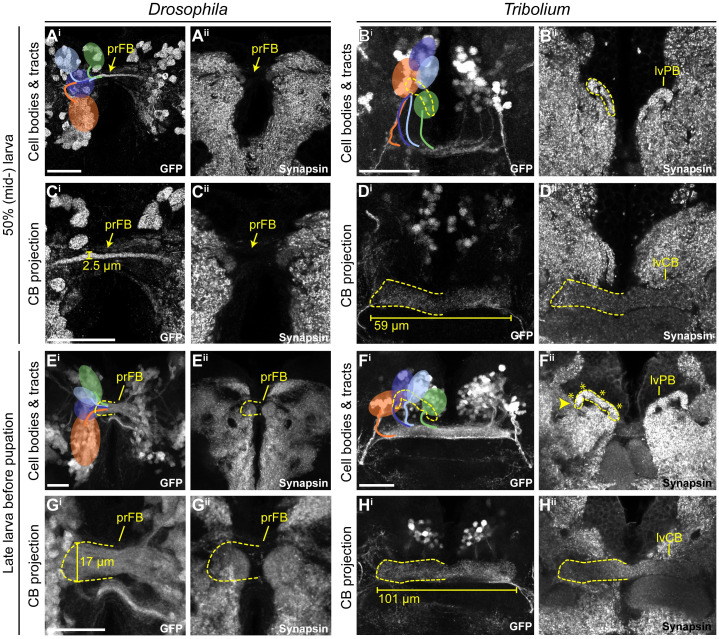
Fig 6. In both species, the rx genetic neural lineage shows substantial growth.
During larval stages, the identified cell clusters and their projections retained their position but proliferated so that larger cell clusters and thicker and larger projections were built. (A-D) Depicted are projections at midlarval stages (50% of larval developmental time) in which cell number and projections have qualitatively increased in number and size, respectively. (E-H) Shown are late larval stages before pupation, in which cell numbers and projection sizes have increased greatly from 50%. The late lvPB of Tribolium can be divided into discrete columns already, indicated by 4 asterisks on one hemisphere. Bars in C, D, G, and H indicate the size increase of midline structures. In Drosophila, the prFB increased in width from 2.5 to 17 μm from 50 to 95% of larval development. In L1, the prFB is nondistinguishable using the rx-GFP line. The central body of the Tribolium L1 brain displayed in Fig 4 was 51.6 μm long, the midlarval lvCB was 58.7 μm, and the late larval lvCB was 100.9 μm long. For Drosophila n-ventral and for Tribolium n-anterior is up (see Fig 4 for details). Scale bars represent 25 μm and apply to panels i and ii and in case of Tribolium to D and H, respectively. GFP, green fluorescent protein; L1, first instar larval; lvCB, larval central body; lvPB, larval protocerebral bridge; n, neuraxis-referring; pr, primordium; rx, retinal homeobox. Original data: https://figshare.com/articles/Fig6/11339813.

