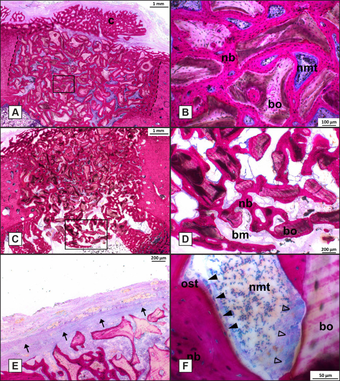
Figure 6.
Histological analysis of bone defects at week 6 (Methylene Blue Basic fuchsin stain, original magnification x400).
A = Histological section of F125 treated defect.
B = High magnification of the boxed area in (A).
C = F125 treated defect showing Bio-Oss® granules extending into the bone marrow.
D = High magnification of boxed area in (C).
E = F20 treated defect showing remnants of the collagen membrane.
F = High magnification of F50 treated defect showing a void where osteoid tissue and osteoblasts lined the newly formed bone and osteoclast-like cells lined the surface of Bio-Oss® granules.
Dashed line = defect border; dashed oval = osteon; nb = new bone; cb = cortical bone; bo = Bio-Oss®; nmt = non-mineralized tissue; closed arrowhead = osteoblasts; bm = bone marrow; ost = osteoid tissue; open arrowhead = osteoclast-like cells.

