Figure 1.

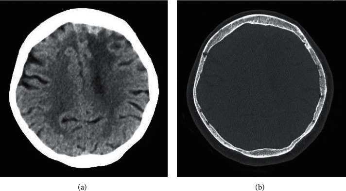
CT head: (a) left frontal extra-axial lesion with patchy calcification and vasogenic oedema; (b) CT bone window setting showing osteopenia but no bony erosion or hyperostosis.

CT head: (a) left frontal extra-axial lesion with patchy calcification and vasogenic oedema; (b) CT bone window setting showing osteopenia but no bony erosion or hyperostosis.