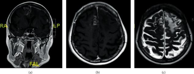
Figure 2.

MRI brain. (a, b) Coronal and axial T1 postcontrast images showing pachymeningeal enhancement in the left frontal region and leptomeningeal enhancement in the left superior and middle frontal sulci, with vasogenic oedema. A nodular leptomeningeal enhancement is noted within the right parafalcine and superior frontal sulci with minimal underlying vasogenic oedema. There was no diffusion restriction. (c) Axial FLAIR image showing the extent of the vasogenic oedema.
