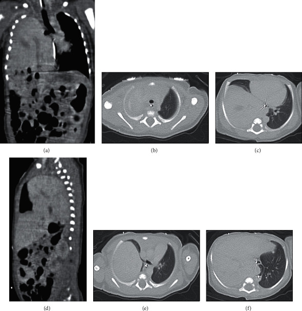
Figure 2.

Computed tomography scans one day after birth. (a) Coronal view of the liver in the right hemithorax extending to the apex with leftward mediastinal shift. (b) Axial images depicting the apex of the lungs, with the right lung apex completely compressed by the liver. (c) Axial view showing the leftward mediastinal shift and the extent of aeration of the right middle lobe. (d) Sagittal view of right lung at the level of greatest aeration. (e) Axial view depicting the right mainstem bronchus and cardiothymic shift. (f) Axial view suggesting possible aplasia of the right lower lobe.
