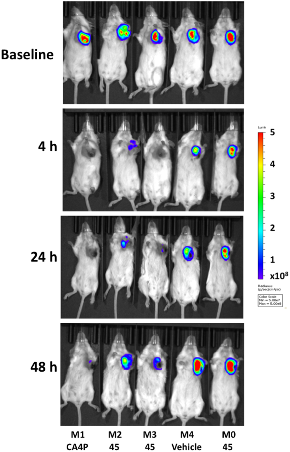
Figure 3.

Bioluminescence images of 4T1-luc tumor-bearing BALB/c mice at various times following VDA administration. Baseline shows mice at 20 min following administration of 120 mg/kg luciferin subcutaneously in the foreback region of five BALB/c mice bearing orthotopic syngeneic 4T1-luc tumors growing in a frontal upper mammary fat pad. Immediately following baseline BLI, mice M2, M3 and M0 were injected IP with 180 mg/kg BAPC 45 dissolved in DPS. M1 received 120 mg/kg CA4P IP and M4 received DPS (vehicle alone). Four, 24 and 48 h later BLI was repeated following administration of fresh luciferin on each occasion. Light emission time courses are presented in Figure 4.
