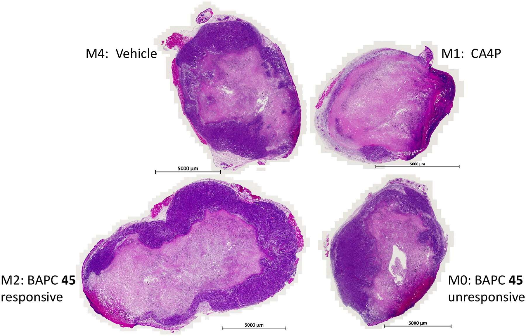
Figure 5.

Histology of 4T1-luc tumors. H&E staining revealed substantial necrosis in all tumors, including the vehicle control. However, necrosis is particularly evident following CA4P (upper right) and in M2, which showed a strong response to BAPC 45. Expanded views of histology are presented in Figure S9 (Supporting Information).
