Fig. 1.

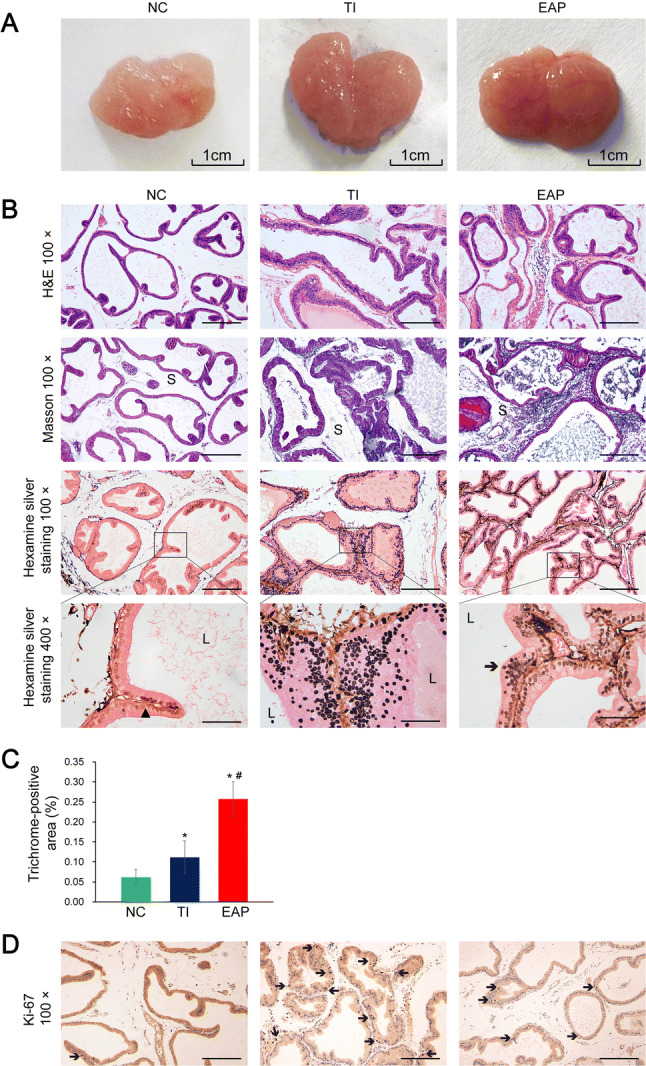
Pathological manifestations in the rat prostate samples. a Appearance of the ventral lobes on both sides of the prostate glands. The scale bar in (a) is mentioned. b H&E staining, Masson’s trichrome staining, and hexamine silver staining of the prostate samples of the rats. The blue colors in the Masson’s trichrome-stained slices indicate collagen fibers. In the hexamine silver-stained slices, the black solid triangular patterns indicate the intact basement membrane and the black arrows indicate the stacking of cells. S stroma, L lumina. c Trichrome-stained areas in the slices (%): the areas positively stained by the Masson’s trichrome stain. d IHC staining for detecting Ki-67 in the rat prostate samples. The black arrows indicate Ki-67-positive cells with proliferative potential. Scale bar, 200 μm under 100× magnification and 50 μm under 400× magnification. Each bar in the graph represents the mean ± S.D. *Significant difference compared to the NC group, P < 0.05; #significant difference compared to the TI group, P < 0.05
