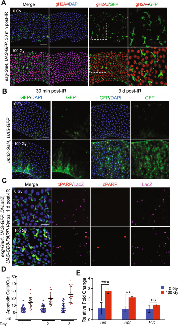
Figure 2.
Exposure to radiation causes DNA damage and cell death in enterocytes and inhibition of ISC proliferation. (A) Midguts were stained with anti-γ-H2Av antibody and DAPI. Guts from esg-Gal4, UAS-GFP flies were dissected 30 min after irradiation with (bottom panels) or without (top panels) 100 Gy. Right panels are magnified images of the white square in the left side panels. Yellow asterisks indicate the both γ-H2Av and GFP positive cells. White asterisks indicate the γ-H2Av negative GFP positive cells. Scale bar indicates 40 µm. (B) Midguts were stained with anti-GFP antibody and DAPI. Guts from upd3-Gal4, UAS-GFP flies were dissected 30 min and 3 days after irradiation with (bottom panels) or without (top panels) 100 Gy. Scale bar indicates 40 µm. (C) Midguts were stained with anti-cPARP antibody, anti-LacZ antibody and DAPI. Guts from esg-Gal4, UAS-GFP, Dl-LacZ, UAS-CD8-PARP-Venus flies were dissected 1 day after irradiation with (bottom panels) or without (top panels) 100 Gy. Yellow asterisk indicates the both cPARP and LacZ positive cell. Scale bar indicates 20 µm. (D) Ethidium bromide-Acridine Orange staining was performed in guts of w1118 female flies after irradiation (100 Gy) at indicated time points. The results are plotted as the mean number of apoptotic cells per gut and presented and mean apoptotic cells and error bars indicate S.D. of 2 independent experiments with at least 10 guts each. (E) Relative fold change in the expression of Hid, Rpr and Puc in the dissected gut 24 h after irradiation. The error bars indicate S.D. of 4 replicates. (***p < 0.001, **p < 0.01, nsp > 0.05 by t-test).

