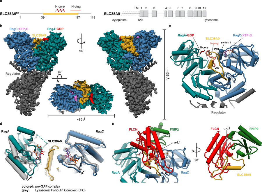
Fig. 2: Cryo-EM structure of the pre-GAP complex.

a, Domain organization of SLC38A9. The construct with structural annotations is shown (left, SLC38A9NT) in context of full-length SLC38A9 (right). Domain boundaries and cellular location of N and C-terminus are indicated. Yellow, resolved in the structure; Dashed box, used construct; TM, transmembrane helix. b, Cryo-EM density of the pre-GAP complex. c, Close-up of the SLC38A9-Rag interaction represented as pipes (α-helices) and planks (β-strands). d, Overlay of Rag GTPases from the pre-GAP complex (colored) with the LFC (grey). e, FLCN:FNIP2 (red, green) from the LFC overlaid onto the pre-GAP complex based on the alignment shown in d, illustrating resulting clashes. Rags are omitted for clarity (right).
