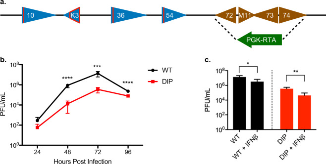
Fig. 1. Construction of DIP virus and its replication properties in vitro.
a Schematic representation of mutations introduced in the MHV-68 genome to generate the DIP vaccine. Red lines indicate insertion of translation stop codons into ORF10, ORF36, and ORF54. The open red tetragon indicates deletion of the coding sequence in K3. The latency locus was replaced by the RTA cassette (arrowhead) constitutively driven by the PGK promoter. b Growth curves of the WT and DIP viruses in 3T3 cells using MOI = 0.01 and measured by plaque assay to quantify virion production. c NIH 3T3 cells were either mock treated or treated with 100 U mL−1 IFN-β for 24 h then infected with either WT or DIP virus at MOI = 0.01 for 72 h. Virion production was quantified with plaque assays. All experiments were performed in triplicate and statistical significance was analyzed by a two-tailed Student’s t-test. P < 0.05*, P < 0.01**, P < 0.001***, and P < 0.0001****. Graphs represent means of triplicates with standard deviations (SD) indicated by error bars.

