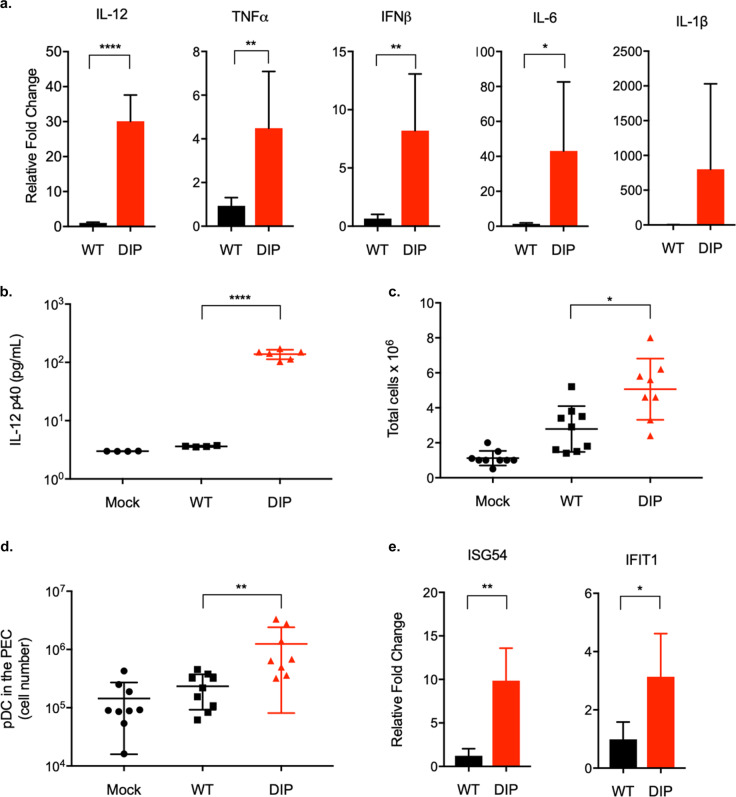
Fig. 7. DIP elicits inflammatory and immunomodulatory cytokines.
Mouse BMDMs were infected with WT or DIP at MOI = 1 (triplicate). a Total RNA was extracted 24 h post-infection for reverse transcription and qPCR to measure the expression levels of IFN-β, IL-1β, TNF-α, IL-6, IL-12, and β-actin. Cytokine RNA expression was normalized against β-actin and the relative fold change was calculated by comparison with mock-infected BMDM. b Supernatants were collected 24 h post-infection to measure IL-12p40 production by ELISA. Mice were either mock-infected or intraperitoneally injected with 105 PFU WT or DIP. PECs were collected at 48 h post-infection. c Total cell numbers in the PECs were counted. d The pDCs were identified by gating on the Lin-(CD3-CD19-NK1.1-)B220+CD11cIntPDCA-1+ population. e Total RNA was extracted from the PECs. RNA expression of ISGs was analyzed by quantitative PCR. Means and SD indicated by error bars were plotted. Statistical significance was analyzed by a two-tailed Student’s t-test. P < 0.05*, P < 0.01**, P < 0.001***, and P < 0.0001****.

