
Fig. 2:
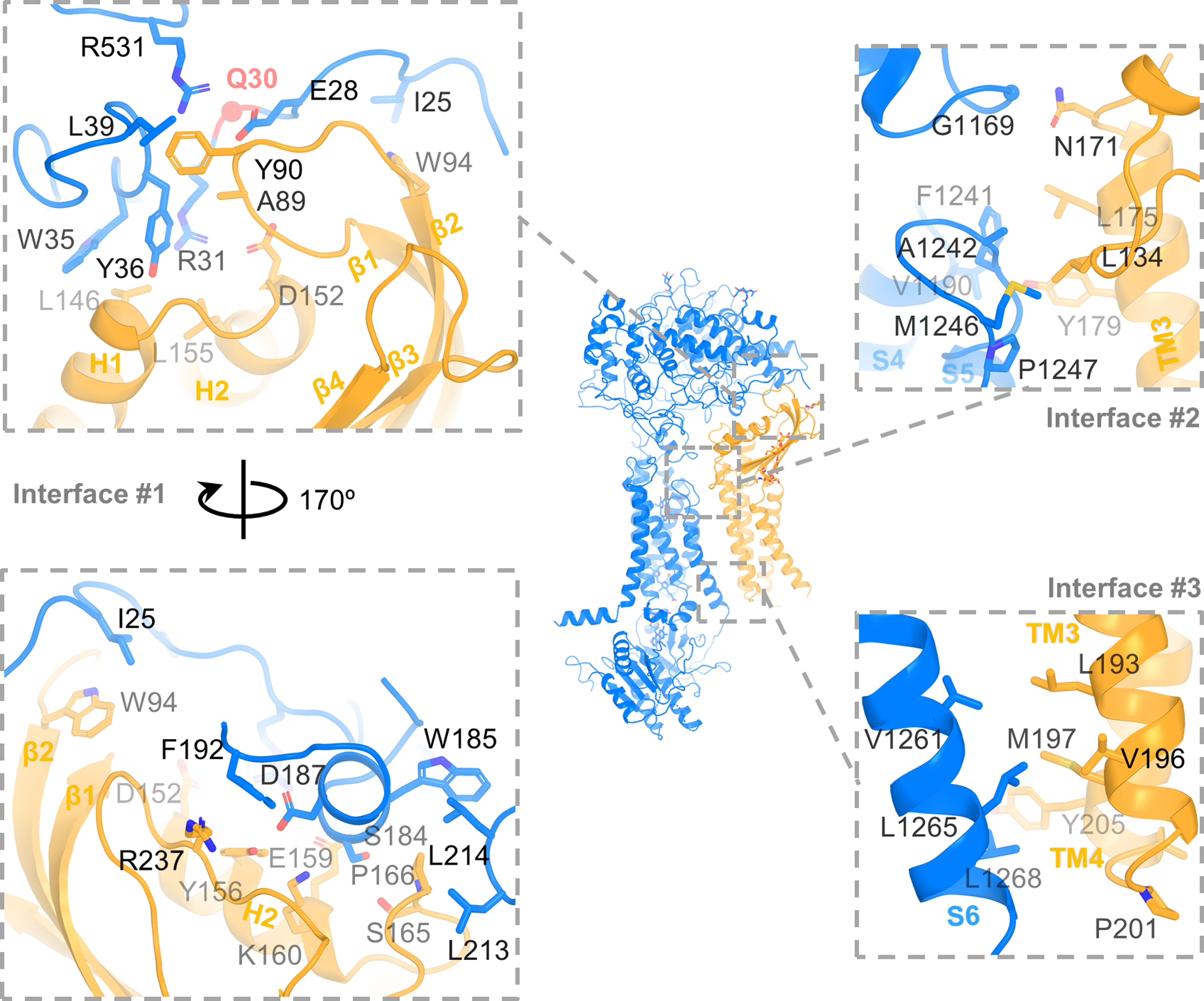
Interaction between DUOX1 and DUOXA1. Three major interfaces between DUOX1 and DUOXA1 are zoomed in and shown in dashed boxes. Left, Interface #1 between extracellular domains with two 170° rotational views. Q30 is shown as a red sphere. Top right: interface #2 at the extracellular side of the cell membrane. Bottom right: interface #3 at the cytosolic side of the cell membrane.