
Fig. 5:
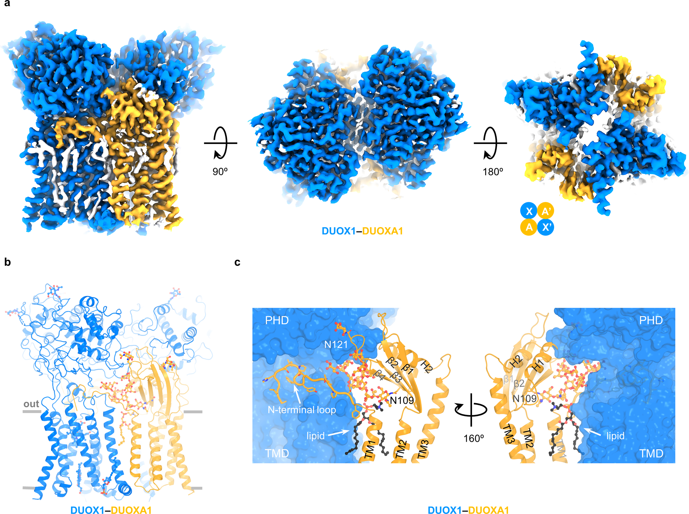
Structure of DUOX1–DUOXA1 in dimer-of-dimer configuration. a, Cryo-EM density map of DUOX1–DUOXA1 in the dimer-of-dimer configuration in three different views. Unexplained or lipid densities are colored in white. The arrangement of the dimer of dimer is depicted using cartoon cycles. b, The structural model of DUOX1–DUOXA1 in the dimer-of-dimer configuration. Sugars, lipids and hemes are shown as sticks and balls. c, Interaction between DUOX1 and DUOXA1. The lipid molecule between DUOX1 and DUOXA1 is colored in black. DUOX1 is presented as surface. TM4 and TM5 of DUOXA1 are not shown for clarity. The N-terminal loop of DUOXA1 is shown as cartoon with side chains.