
Extended Data Fig. 2. Structure of the PHD of DUOX1.
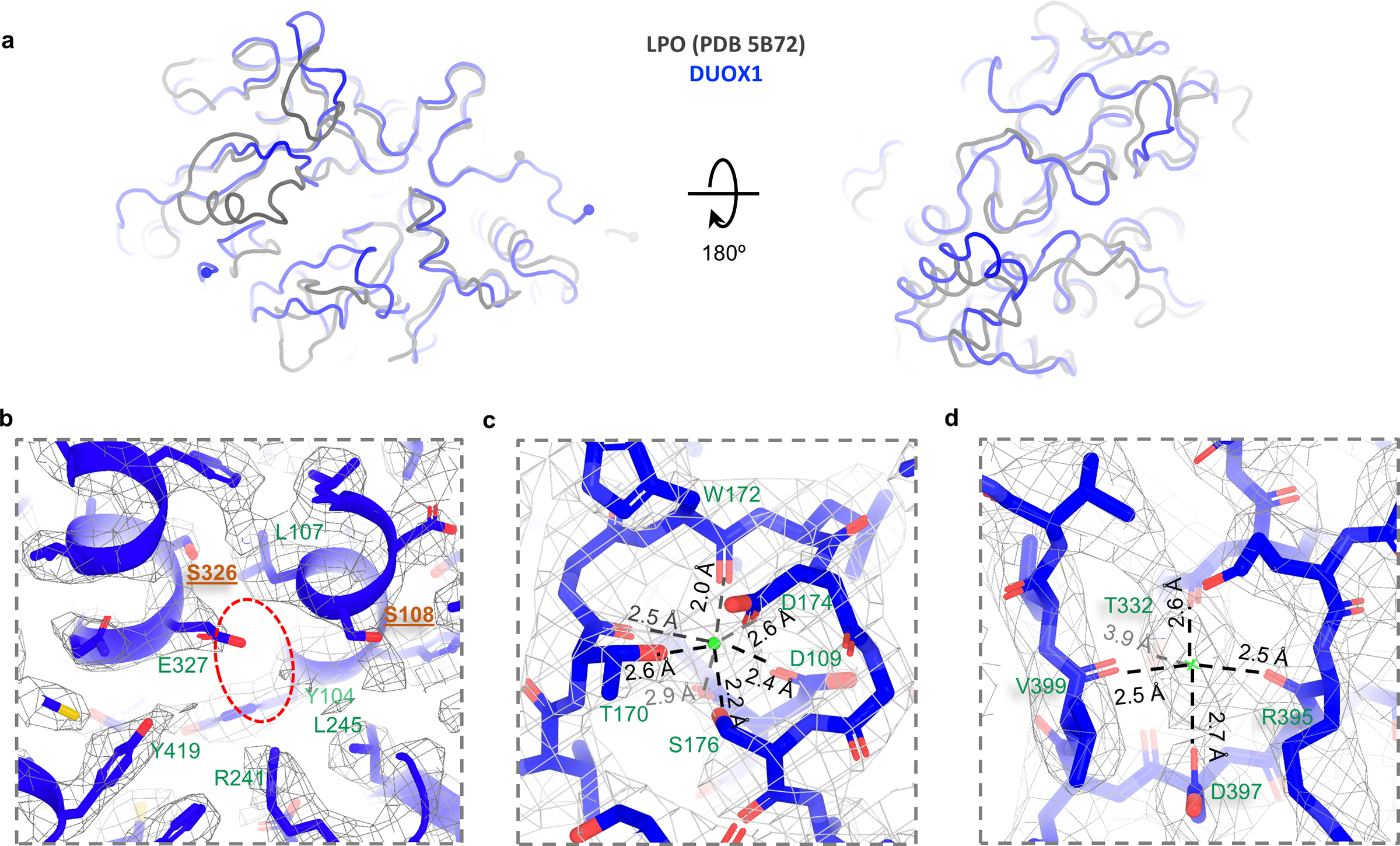
a, Superimposition between LPO and PHD of DUOX1. LPO and DUOX1 are colored in grey and blue, respectively. b, Putative heme-binding site. The possible heme-binding pocket indicated by a red oval. Positions of Ser326 and Ser108 (colored in brown) are where the heme-coordinating histidines are located. c, The putative calcium-binding site and surrounding residues. Calcium is indicated by a green sphere. d, A potential ion binding site and surrounding residues. Cryo-EM density is contoured by grey meshes.