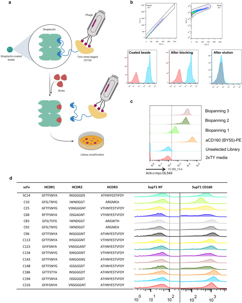
Figure 2.
Biopanning of the scFv phage display library on Streptactin magnetic beads. (a) Schematic representation of the biopanning process employing the StreptagII-Streptactin system. CD160 fused with Twin streptag was capture directly from cell supernatant on the surface of Streptactin magnetic beads. Phages were incubated with the antigen’ coated beads and bound phages were subsequently recovered by eluting with a 30 mM biotin solution and used to amplify the library for the next biopanning round. (b) The process of coating and elution of the beads throughout the biopanning process was assessed by flow cytometry analysis. These were stained during the several steps of the procedure (coating, blocking with 3%BSA and elution) using commercial anti CD160-PE. (c) Enrichment of the phage library for CD160 was determined after three rounds of biopanning on beads. The bacterial library from the different rounds of selection was induced to express the soluble scFv fragment in the supernatant. This was used to stain SupT1 cells overexpressing human CD160, due to the presence of a myc-tag at the C-terminus of productive scFvs we were able to detect binding using anti myc antibody. 2xTY media + anti myc-tag-DL549 and the commercial anti-CD160-PE Ab were used as controls. (d) The 15 individual bacterial colonies identified in the third round of biopanning carrying unique combination of CDR1,2 and 3 were screened by against SupT1 CD160 positive and negative cells (NT).

