Figure 6.
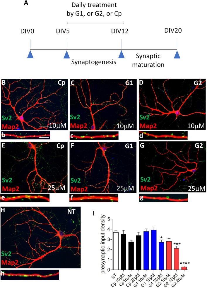
Inhibiting 3S-HS by G2 peptide impairs synapse formation. (A) graphical sketch of the daily treatment (DIV 5 to DIV 12) of immature primary hippocampal neurons in culture with G1, G2, or control Cp peptides. (B–G) co-immunofluorescent neurons labelled with Map2 (red) and Sv2 (green) after treatment with peptides at 10 µM or 25 µM, and H, in non-treated cell cultures (NT). A high magnification of a dendrite bearing Sv2-immunoreactive puncta is shown for each condition (b–h). (I) quantification of the density of presynaptic Sv2-immunoreactive puncta (n = 2 independent experiments). Dunn’s selected comparison test: G2, 25 µM versus NT, ****p < 0.0001; G2, 15 µM versus NT, ***p = 0.0006; G1, 50 µM versus NT, *p = 0.0114. Bar in (B), 75 µm (applies for B–H); Bar in (b), 5 µm, (applies for b–h).

