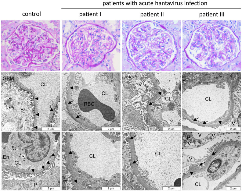
Figure 1.
Light and electron microscopy of the glomerular filtration barrier in three patients with acute hantavirus infection and one healthy control. The glomerular filtration barrier of three patients with acute hantavirus infection (Heidelberg biopsy register) and one healthy control (living kidney donation) were analyzed by HE staining and transmission electron microscopy. Arrowheads indicate intact podocyte foot processes; arrows show effacement of podocyte foot processes. CL = capillary lumen, En = endothelium, GBM = glomerular basement membrane, P = podocyte, RBC = red blood cell, V = vacuole.

