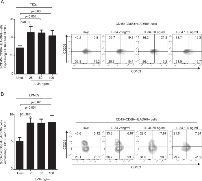
Fig. 6. IL-34 enhances the expression of markers of type 2-polarized macrophages.
A, TICs (A) LPMCs (B) were either left unstimulated (Unst) or stimulated with increasing doses of human IL-34 (25–100 ng/ml) for 24 h, and the percentages of live CD45 + , CD68 + ,HLA-DRII + cells expressing CD163 and CD206 were analyzed by flow cytometry. Data are expressed as mean ± SEM of seven experiments. Right panels: representative dotplots showing CD163 and CD206 expression in live CD45 + CD68 + HLA-DRII + TICs (A) or LPMCs (B) isolated from respectively tumoral and non-tumoral samples of one patient with colon rectal cancer (CRC) and treated as described above.

