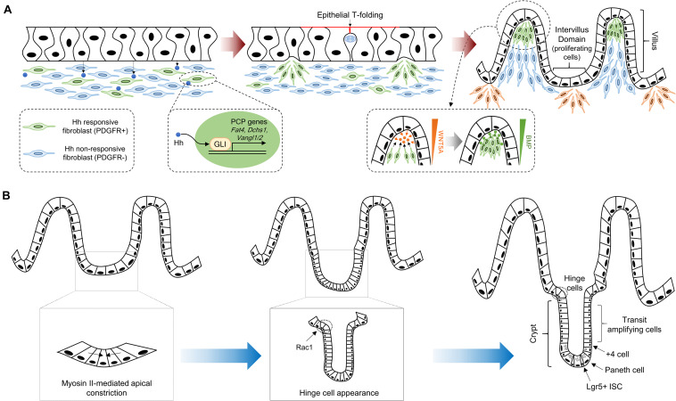
FIGURE 1.
Crypt-villus morphogenesis during mouse development. (A) The pseudostratified intestinal epithelium secretes Hh ligands that induce expression of PCP genes, such as Fat4, Dchs1, Vangl1, and Vangl2, via GLI transcriptional factor activation. Clustered mesenchymal cells (green color) cause deformation of intestinal epithelium through apical membrane invagination with the emergence of epithelial T-folding. The clustered mesenchymal cells underneath the tip of the villus, which are formed by directional migration toward chemoattractants such as WNT5A, enforce villification and differentiation of intestinal epithelium via secretion of soluble ligands including BMPs. New mesenchymal clusters (orange color) beneath the intervillus domain lead to the second round of villification processes. (B) Intervillus proliferating cells transform into crypts. The initiation of crypt formation is driven by myosin II-mediated apical constriction and invagination of the crypt cells. Subsequently, hinge formation in the crypt neck is initiated to compartmentalize crypts and villus, and Rac1 controls this process through the wedge-shaped hinge cell formation. These crypts contain Lgr5 + ISCs intercalated between Paneth cells at the crypt base, and transit amplifying cells.

