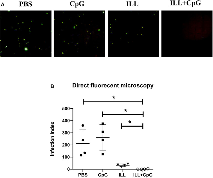
Figure 4.
Parasite burden assay in popliteal lymph node using direct fluorescent microscopy. Eight weeks after challenge with L. majorEGFP, mice were sacrificed and popliteal lymph nodes were isolated. (A) The cell suspension was prepared. The parasites expressing green fluorescent protein (GFP) were imaged using a fluorescent microscope. (B) To compare parasite burden between groups, the infection index was calculated by multiplying the percentage of infected macrophages by the average number of parasites per cell. The infection index was significantly lower in immunized groups than controls. To compare the means, the Mann–Whitney U-test was used. (* P ≤ 0.050). Data are shown as mean ± SD.

