FIGURE 4.
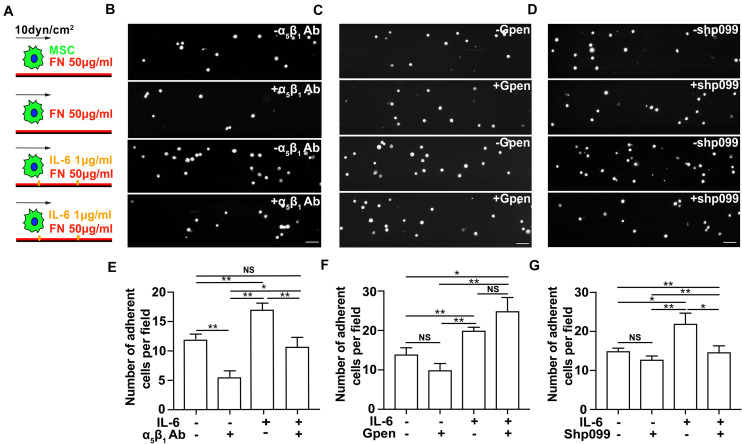
The interleukin-6 (IL-6)-dependent adhesion enhancement on mesenchymal stem cell (MSC) adhesion under shear is mediated through SHP2–integrinα5β1 signaling axis. Schematics (A) of MSC perfusion over microfluidic channels coated with 50 μg/ml of fibronectin (FN) in the absence (first and second rows in B–D) or in the presence of 1 μg/ml IL-6 (third and fourth rows in B–D), of which are their representative images of attached MSCs pretreated with α5β1 blocking mAb (B), Gpen (C), or shp099 (D) for 30 min before perfusion and are corresponding average numbers of adherent MSCs per field (E–G), respectively. Scale bars in panels (B–D) refer to 200 μm. **, *, and NS in panels (E–G) refer to p < 0.01, p < 0.05, and no significance, respectively. Error bars in panels (E–G) represent SEM of three repeats.

