Figure 1.
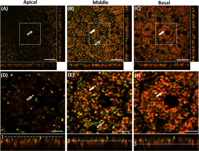
Confocal image planes of the RPE mosaic of Abca4−/− mouse acquired at different depths with a 561-nm laser. The image strips at the bottom and right side of each (x, y) panel show the side views of the confocal three-dimensional image stack (x,z) and (y,z), respectively. (A) Apical side of RPE, (B) mid-RPE region, and (C) basal part of the RPE confocal image stack. (D–F) Enlarged images of the regions indicated by the white-dashed square boxes in A–F, respectively. Hollow white arrows indicate melanosomes, and solid white arrows indicate putative lipofuscin granules. White dashed lines in the bottom panel (1–3) show the position of the selected planes in the RPE volume: (1) apical, (2) middle, and (3) basal. Scale bars: 20 µm (A–C), 10 µm (D–F).

