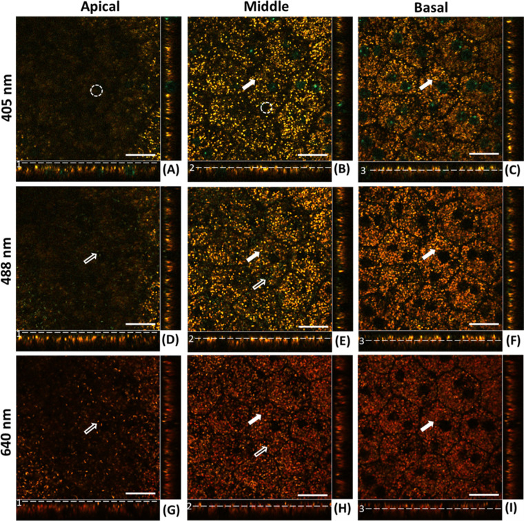
Figure 2.
Confocal image planes of the RPE mosaic of Abca4−/− mouse acquired with 405-nm, 488-nm, and 640-nm, excitation wavelengths, respectively. (A, D, G) Apical plain of RPE. (B, E, H) Mid-RPE region plane. (C, F, I) Basal plane of RPE. Image strips at the bottom and right side of each (x, y) panel show the side views of the confocal three-dimensional image stack (x,z) and (y,z), respectively. White solid arrows indicate the lipofuscin granules appearing in the middle and basal plane of the RPE. Hollow arrows indicate the melanosomes. Dashed white circles highlight the lack of fluorescence of the melanosomes when excited at 405 nm. Scale bars: 20 µm.

