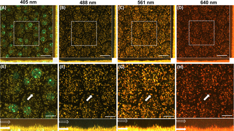
Figure 3.
Confocal images of RPE cells of an albino mouse (melanin free). Images acquired with excitation wavelengths of (A) 405 nm, (B) 488 nm, (C) 561 nm, and (D) 640 nm. (E–H) Magnified image of the dashed box shown in A–D. Hollow white arrows (in bottom panel) indicate the apical part of the RPE. Solid white arrows indicate representative lipofuscin granules in the basal part of the RPE. Scale bars: 20 µm (A–D), 10 µm (E–H).

