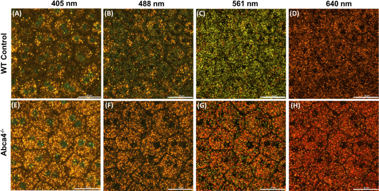
Figure 5.
Confocal images (depth intensity projection) of the RPE cells mosaic from flat-mount of WT control (A–D) and Abca4−/− (E–H) for the excitation wavelengths of 405 nm (A, E), 488 nm (B, F), 561 nm (C, G), and 638 nm (D, H). There are a decreased number of melanosomes (green) and an increased density of lipofuscin granules (yellow-orange-red) in Abca4−/− as compared to WT. Scale bars: 25 µm.

