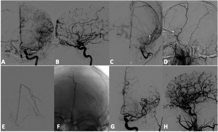
Figure 1.
Patient Nr 9. Preliminary DSA during selective injection of left internal and external carotid arteries (a, b, c, d) shows a DAVF of the Crista Galli with pial reflux in right cerebral hemisphere. Left MMA was the selected way for the embolization but the microcatheter stopped in the basal frontal branch (arrows). Frontal angiogram (e) during superselective injection through the microcatheter in the basal branch of the left MMA. Unsubtracted image (f) shows SQUID 12 cast at the end of the endovascular treatment. Despite the distance between the microcatheter and the DAVF, injection time was 25 minutes and the amount of embolic agent was 4,5 ml. Final angiograms of the left CCA (g, h) demonstrates the complete occlusion of the cDAVF.

