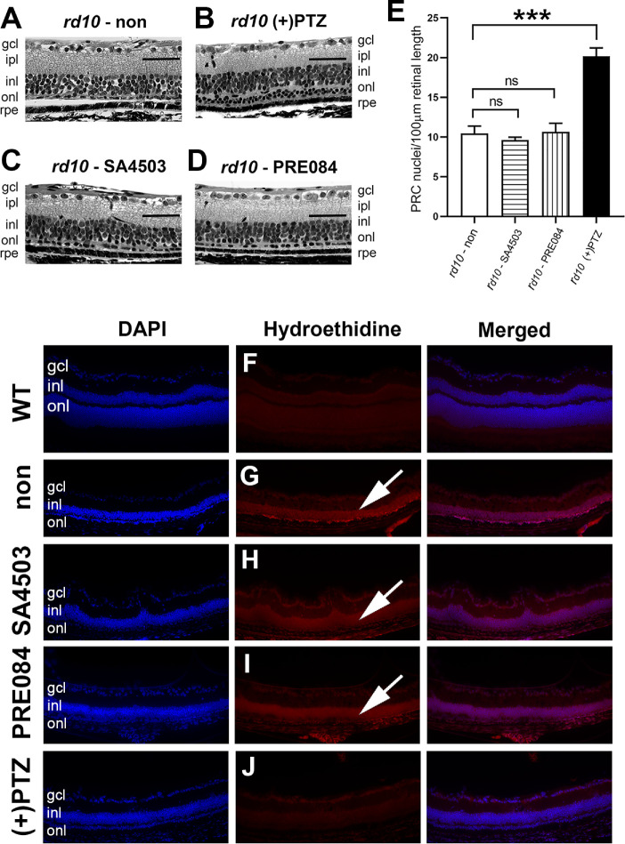
Figure 8.
Assessment of retinal histologic structure in rd10 mice treated with SA4503, PRE084, or (+)-PTZ. Rd10 mice were administered SA4503, PRE084, or (+)-PTZ every other day beginning at P14. Mice were euthanized at P42 and eyes prepared for histology. Representative photomicrographs of hematoxylin-eosin stained retinal sections are provided for (A) rd10 (nontreated), (B) rd10+PTZ, (C) rd10 - SA4503, and (D) rd10 - PRE084. (E) The number of PRC nuclei within the ONL were counted and data expressed per 100 µm retinal length. One-way ANOVA, significance is depicted as ***P < 0.001, ns = not significant. Data are the mean ± SEM of analyses in four to six mice per group. (Arrows in A–D point to row[s] of PRC nuclei.) (F–J) Immunodetection of hydroethidine, which fluoresces red upon reaction with superoxide species in retinal cryosections of (F) WT, (G) rd10-non, (four to six) rd10-SA4503, (I) rd10-PRE084, and (J) rd10-(+)-PTZ. (Arrows in G–I point to red fluorescence.) gcl, ganglion cell layer; ipl, inner plexiform layer. Calibration bars (A–D) = 50 µm; (F–J) = 100 µm.

