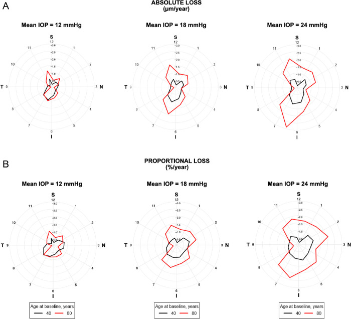
Figure 4.
Polar plots illustrating the estimated rates of change in RNFL thickness according to the sectors around the optic disc for subjects 40 and 80 years of age and different levels of average IOP during follow-up. It can be seen that the impact of IOP was significantly greater in older eyes compared to younger ones and that the effect was markedly greater in the inferior and superior sectors of the optic nerve, in terms of both absolute loss (µm/year) (A) and percentage of loss from the baseline thickness (%/year) (B) for each sector.

