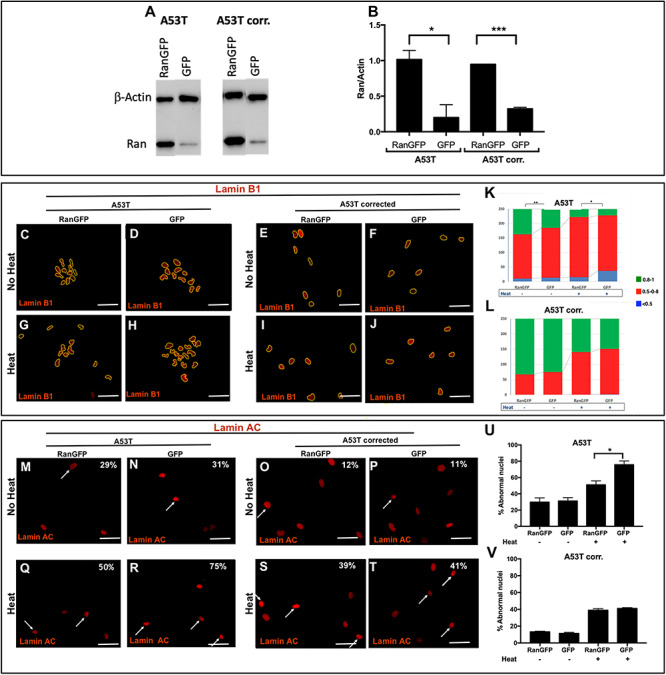
Figure 4.

The effects of RAN overexpression on the integrity and function of the nuclear envelope. A53T hiPSC-derived MD NPCs and the isogenic corrected control were transduced with pLenti-IRES-GFP and pLenti-RAN-IRES-GFP. (A) Overexpression of RAN was confirmed by western blot analysis using anti-RAN antibody (25 kDa) normalized to β-actin (42 kDa). (B) RAN protein levels were quantified in two independent western-blot experiments. Higher levels of RAN protein were detected in the A53T hiPSC-derived MD NPCs and the isogenic corrected control. (C-V) The effect of RAN overexpression on the integrity of the nuclear envelope and its sensitivity to heat-shock was evaluated by immunocytochemistry for Lamin B1 (C-J) and Lamin A/C (M-T) in the A53T hiPSC-derived MD NPCs and the isogenic corrected control in regular conditions (C-F, M-P) and upon heat-shock (G-J, Q-T). The overexpression of RAN was visualized using GFP reporter; thus, for these set of experiments, we used red fluorescent conjugated secondary antibody to detect the staining of Lamin B1 (C-J) and Lamin A/C (M-T). Percentages indicate the proportion of cells with folded/blebbed nuclear morphology, white arrows denote abnormal nuclei (M-T). (K, L) The nuclear circularity data are plotted as frequency distributions of for 250 cells (n = 2). (U, V) Percentages indicate the proportion of cells with folded/blebbed nuclear morphology. Bars represents mean ± SEM, n = 2. (K, L) *P < 0.05, **P < 0.001, Kolmogorov–Smirnov test; (B, U, V) *P < 0.05, ***P < 0.001 Student’s t-test. Scale bars: 10 μm.
