
Figure 5.
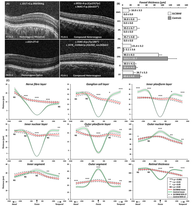
(A) Foveal tomograms associated with different SLC38A8 mutations. There is no evidence of a foveal pit or photoreceptor OS lengthening. In F1:II-1, F2:II-1 and F7:II-3, there some evidence of ONL widening, consistent with grade 3 foveal hypoplasia. In F8:II-1, there is no ONL widening which is characteristic of grade 4 foveal hypoplasia. (B) Bar graph showing the mean thickness for intra-retinal layers at the fovea for SLC38A8 patients and controls. The error bars indicate SD. (C) Mean thickness plots of each retinal layer with 95% confidence intervals for SLC38A8 patients (red) and controls (green). X-axis represents distance away from fovea in the nasal and temporal directions (in microns). For statistical comparisons thickness measurements were compared at the fovea, 1 and 2 mm from fovea in both the nasal and temporal directions. Levels of significance indicated alongside the relevant position (*** = P < 0.001; ** = P < 0.01; * = P < 0.05; NS = not significant (P > 0.05)). Abbreviations: RNFL = retinal nerve fibre layer; GCL = ganglion cell layer; IPL = inner plexiform layer; INL = inner nuclear layer; OPL = outer plexiform layer; ONL = outer nuclear layer; IS = inner segment; OS = outer segment.