
Figure 7.
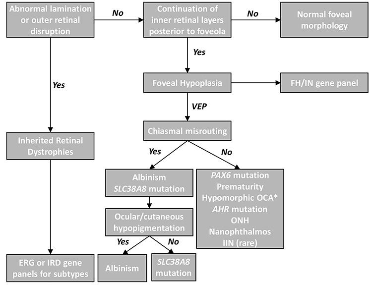
Clinical diagnostic algorithm for abnormal retinal development. The hallmark of typical foveal hypoplasia is continuation of inner retinal layers posterior to the foveola. Outer retinal changes or abnormal lamination suggests an inherited retinal dystrophy (IRD). Electroretinogram (ERG) or IRD gene panels can differentiate between the IRD subtypes. FH or IN gene panels can readily differentiate the genetic subtypes that can cause FH. In the absence of genetic panels, VEP is helpful in broadly differentiating the diagnoses based on chiasmal misrouting. In some cases of albinism* (hypomorphic mutations or carriers), normal VEP responses maybe present. ONH = optic nerve hypoplasia, IIN = idiopathic infantile nystagmus.