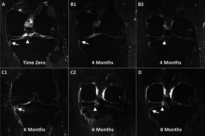
Figure 2.
Serial magnetic resonance imaging showing the progression of a meniscotibial (MT) ligament tear, medial meniscal extrusion, and eventual medial meniscus posterior root tear. (A) Initial T2-weighted coronal imaging demonstrates bright MT ligament edema (arrow) while the root remains intact (arrowhead). There is a degenerative signal in the meniscus with irregularity and trace extrusion. (B1) Repeat imaging at 4 months demonstrates a further increase in the signal and MT ligament attenuation (arrow), increased extrusion, and (B2) new partial tearing and an increased signal in the meniscus root (arrowhead). (C1) Imaging at 6 months from baseline demonstrates marked attenuation of the MT ligament (arrow), substantial extrusion, the progression of femoral chondromalacia, and (C2) a concurrent full-thickness radial root tear (arrowhead). (D) At 8 months, there is radiographic loss of the MT ligament (arrow) and clearly visible complete tearing and displacement of the meniscus root (arrowhead).

