
Figure 1.
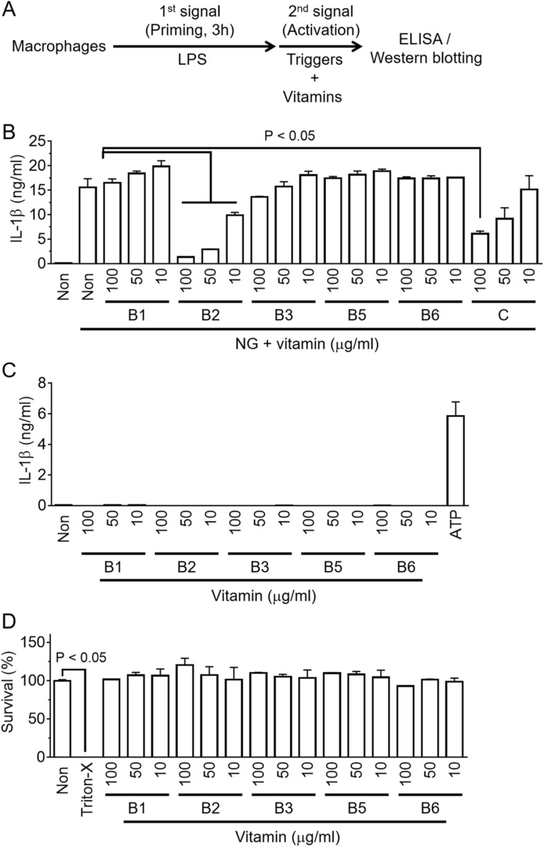
Effects of vitamin B on NLRP3 inflammasome activation. (A) Schematic diagram of the experimental process of inflammasome activation. Macrophages, such as BMDMs or PMA-treated THP-1 cells, were primed with LPS for 3 h, and the inflammasome activation was then triggered in the presence of a vitamin. The indicators of the inflammasome activation were analyzed by ELISA or Western blotting. (B) LPS-primed BMDMs activated the NLRP3 inflammasome with NG with/without a vitamin as indicated. The secretion of IL-1β was measured by ELISA. (C) LPS-primed BMDMs were treated with a vitamin, as indicated without the inflammasome triggers. The ATP treated group was used as a positive control. (D) LPS-primed BMDMs were treated a vitamin, as indicated for 24 h, and the cytotoxicity was analyzed. The survival rates of the non-treated cells were set to 100%, and the cells treated with triton X-100 (Triton-X, 0.01%) were regarded as 0% viability. The bar graph presents the mean ± SD with at least two independent experiments.