Figure 2.
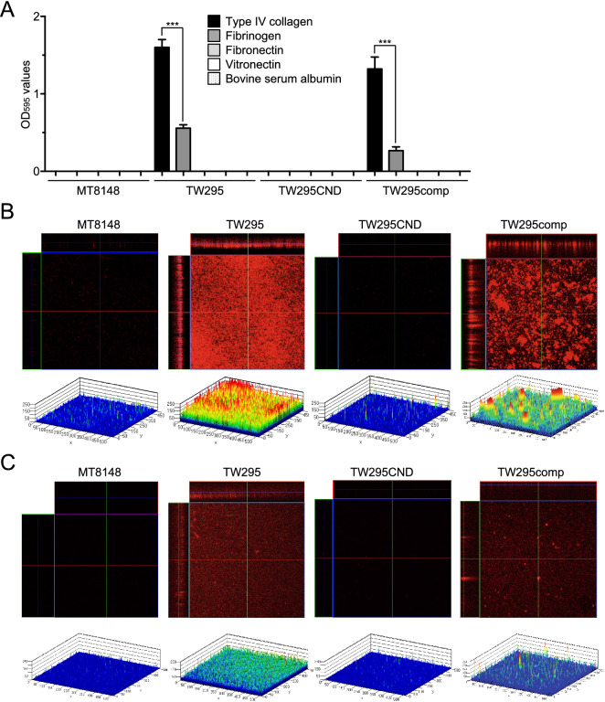
ECM binding of S. mutans strains. (A) Binding of the indicated S. mutans strains to type IV collagen, fibrinogen, fibronectin, or vitronectin. Data are presented as the means ± SD of three technical replicates. ***p < 0.001 by ANOVA followed by Bonferroni’s post hoc test. (B, C) Confocal laser scanning microscopy images (upper panels) and schematics (lower panels) of S. mutans binding to type IV collagen (B) or fibrinogen (C). Color coding of the biofilm thickness: 0–50 µm, blue; 50–100 µm, light blue; 100–150 µm, green; 150–200 µm, yellow; and 200–250 µm, red. All confocal laser scanning microscope images and schematics were made using LSM510.

