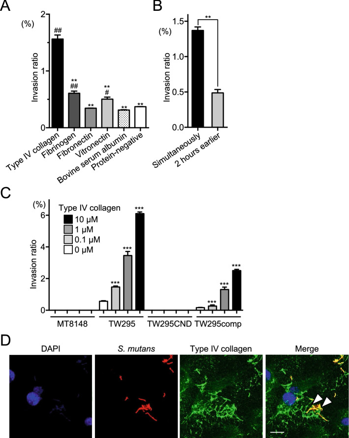
Figure 3.
Invasion of HUVECs by S. mutans in the presence of purified ECM proteins. (A) Invasion ratios of HUVECs after their incubation with S. mutans strain TW295 for 2 h at a multiplicity of infection of 100 in the presence of type IV collagen (140 ng/ml), fibrinogen (2 mg/ml), fibronectin (0.2 mg/ml), vitronectin (500 µg/ml), or bovine serum albumin (1 mg/ml). Data are presented as the means ± SD of three technical replicates. *p < 0.05, **p < 0.01 versus TW295 and #p < 0.05, ##p < 0.01 versus the protein-negative control, using ANOVA followed by Bonferroni’s post hoc test. (B) Ability of TW295 to invade HUVECs when type IV collagen and bacteria were added simultaneously or when type IV collagen was added 2 h before the bacterial infection. Data are presented as the means ± SD of three technical replicates. **p < 0.01 using a Student’s t-test. (C) HUVEC invasion ratio (as described for A) of the indicated strains in the presence of various concentrations of type IV collagen. Data are presented as the means ± SD of four technical replicates. ***p < 0.001 versus no collagen using ANOVA followed by Bonferroni’s post hoc test. (D) Representative confocal laser scanning microscopy images of S. mutans TW295 invading HUVECs. Bacteria are stained red (Alexa Fluor 555-conjugated anti-Cnm antibody), type IV collagen is stained green (Alexa Fluor 488), and nuclei are stained blue (DAPI). Arrowheads indicate areas were type IV collagen and bacteria colocalize. Scale bar, 10 µm. All confocal laser scanning microscope images were taken using LSM510.

