Figure 6.
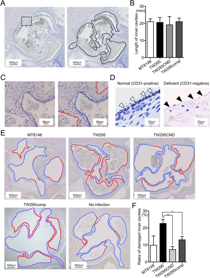
Evaluation of vascular endothelial cell damage in a rat IE model. (A) Representative IHC images of anti-CD31-stained sections of extirpated heart valves from rats infected with S. mutans strain TW295. Black lines indicate the total length of the endocardium. (B) Total length of the endocardium of extirpated hearts from rats infected with the indicated S. mutans strain. Data are presented as the mean ± SD of six biological replicates per strain. (C) Magnified images of the box outlined in (A). Right panel shows endothelial cell damage. Blue and red lines indicate non-damaged (CD31-positive) and damaged (CD31-negative) endocardium, respectively. (D) Representative magnified images of normal (CD31-positive; arrowheads in left panel) and deficient (CD31-negative; arrowheads in right panel) endocardium. (E) Representative images of vascular endothelial cell damage in hearts from rats infected with the indicated S. mutans strains. Blue and red lines indicate non-damaged (CD31-positive) and damaged (CD31-negative) endocardium, respectively. (F) Rates of damaged endocardium following infection by each S. mutans strain. Data are presented as the mean ± SD of six biological replicates per strain. *p < 0.05, **p < 0.01 using ANOVA followed by Bonferroni’s post hoc test. The lengths of total, CD31-positive, and CD31-negative endocardium in the heart valves were measured using WinROOF Vol 5.0 software (Mitsuya Co., Ltd., Tokyo, Japan).

