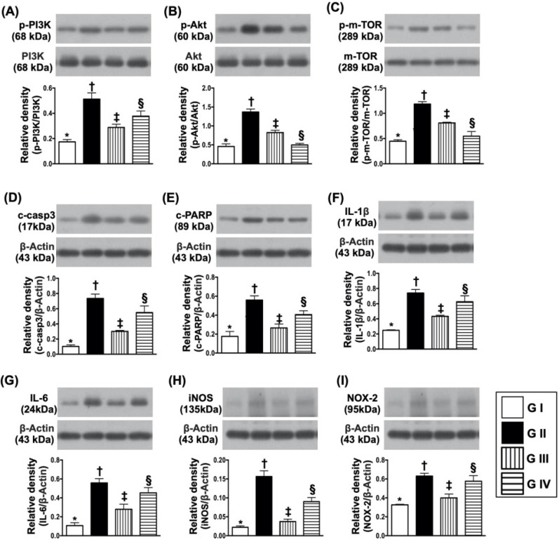
Figure 9.
Protein expressions of cell survival/death signaling, oxidative-stress and inflammatory biomarkers. (A) Protein expression of phosphorylated (p)-PI3K, * vs. other groups with different symbols (†, ‡, §), p<0.001. (B) Protein expression of p-AKT, * vs. other groups with different symbols (†, ‡, §), p<0.0001. (C) Protein expressions of p-m-TOR* vs. other groups with different symbols (†, ‡, §), p<0.0001. (D) Protein expression of cleaved caspase 3 (c-casp3), * vs. other groups with different symbols (†, ‡, §), p<0.0001. (E) Protein expression of c-PARP, * vs. other groups with different symbols (†, ‡, §), p<0.001. (F) Protein expression of interleukin (IL)-1ß, * vs. other groups with different symbols (†, ‡, §), p<0.0001. (G) Protein expression of IL-6, * vs. other groups with different symbols (†, ‡, §), p<0.001. (H) Protein expression of inducible nitric oxide synthase (iNOS), * vs. other groups with different symbols (†, ‡, §), p<0.0001. (I) Protein expression of NOX-2, * vs. other groups with different symbols (†, ‡, §), p<0.003. All statistical analyses were performed by one-way ANOVA, followed by Bonferroni multiple comparison post hoc test (n=4 for each group). Symbols (*, †, ‡, §) indicate significance (at 0.05 level). Group I = Na2 cell only (i.e., control group); Group II = N2a cell treated by H2O2; Group III = N2a cell treated by H2O2 and sitagliptin; Group IV = N2a cell + H2O2 + sitagliptin + LY-294002.

