Fig. 1.
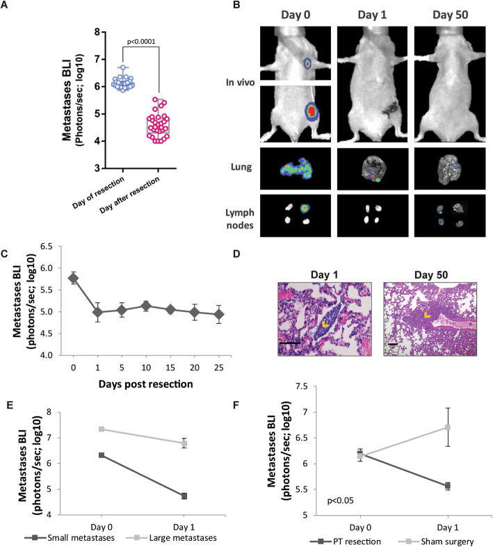
Excision of the primary tumor elicits regression of early-stage metastases. a In vivo quantification of lung and lymph node metastasis by bioluminescence imaging (BLI) immediately before (day 0) and after primary tumor (PT) resection (day 1) (n = 29). Whiskers represent the min and max points. b Representative images of lung and lymph node (LN) metastases, in vivo and ex vivo at day 0 and post-operative days 1 and 50, following tumor excision. c In vivo BLI of metastases over time (n = 6). d Representative images of H&E staining of the lung sections at post-operative days 1 and 50. Orange arrows indicate micro-metastases. Scale bar day 1 = 100 μm, scale bar day 50 = 225 μm. e In vivo quantification of lung and LN metastases by BLI before (day 0) and after PT resection (day 1) in mice bearing small (n = 7) or large (n = 9) metastases. f In vivo quantification of lung and LN metastases by BLI before (day 0) and after (day 1) sham surgery or PT resection. The detection threshold for all BLI figures is ~ 104 photons/s (i.e., log10 = ~ 4). Error bars in c, e, and f represent mean ± SE

