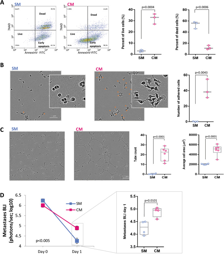
Fig. 2.
CM effects on pro-metastatic processes and metastasis. a Representative images and quantification of flow cytometry for AnnexinV and 7AAD of MDA-MB-231HM cells that were grown in serum-free medium (SM) or MDA-MB-231HM tumor cell-conditioned medium (CM). b Representative images and quantification of adhered cancer cells incubated with SM or CM. Orange arrows mark adhered cells. Scale bar, 100 μm. c Representative images and quantification of tube formation by human endothelial cells incubated with SM or CM on a layer of basement membrane extracellular matrix. Scale bar, 100 μm. d In vivo quantification of lung and LN metastases by BLI before (day 0) and after PT resection (day 1) in mice that received SM or CM, simultaneously with tumor excision (n = 4 per group). The detection threshold is ~ 104 photons/s (i.e., log10 = ~ 4). Whiskers in c and d represent the min and max points. Error bars in a–d represent mean ± SE

