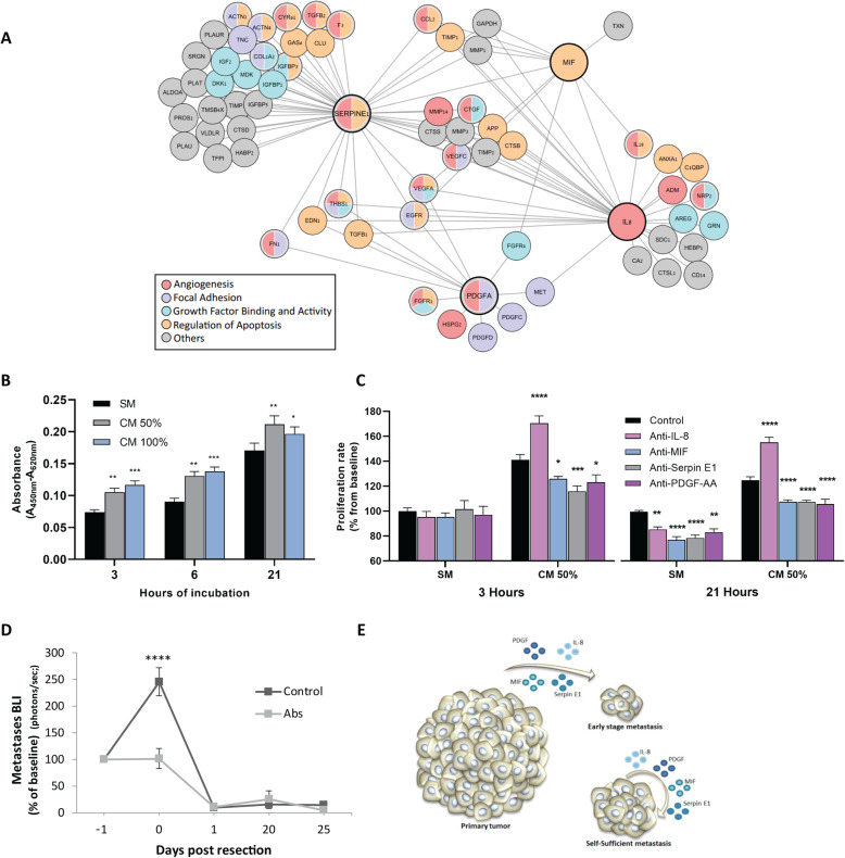
Fig. 3.
Secreted factors and pathways potentially underlying metastatic regression. a Proteomic and GO enrichment analysis identified a network of proteins and enriched pathways. Proteins in this scheme are those connected to at least one of the 4 key factors (denoted by a thick line). b Proliferation of MDA-MB-231HM cells that were cultured in serum-free medium (SM) or in 100% or 50% MDA-MB-231HM tumor cell-conditioned medium (CM), measured after 3, 6, or 21 h of incubation (3 h, n = 20 per media type; 6 and 21 h, n = 16 per media type). Asterisks represent a significant result relative to the SM group in each time point. c Proliferation of MDA-MB-231HM cells that were cultured for 3 or 21 h in SM or CM 50% with IgG isotype control or antibodies against either IL-8, PDGF-AA, Serpin E1, or MIF. Data is presented as percent from IgG control SM in each time point (each antibody, n = 4; IgG control, n = 16). Asterisks represent a significant result relative to the control group. d In vivo long-term quantification of metastasis following cocktail administration of the four neutralizing antibodies (IL-8, MIF, PDGF-AA, and Serpin E1; n = 4) or IgG control (n = 5). e A scheme of the hypothesized model based on our results, suggesting that primary tumor secretome is crucial for the survival of early-stage micro-metastases, but not for larger metastases. Abs, antibodies. Error bars in b–d represent mean ± SE. ****p < 0.0001, ***p < 0.001, **p < 0.01, *p < 0.05

