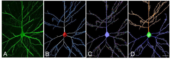
Figure 5.
(A) Confocal microscopy image of an intracellularly injected human layer III pyramidal neuron. (B) Three-dimensional reconstruction of the morphology of the cell shown in (A), using Imaris. (C) The building of the soma, dendrites, and spines from the neuron shown in (B), using Neuronize. (D) Neurolucida visualization of the neuron shown in (C). Scale bar (in D): 20 μm in (A–D).

