
Figure 6.
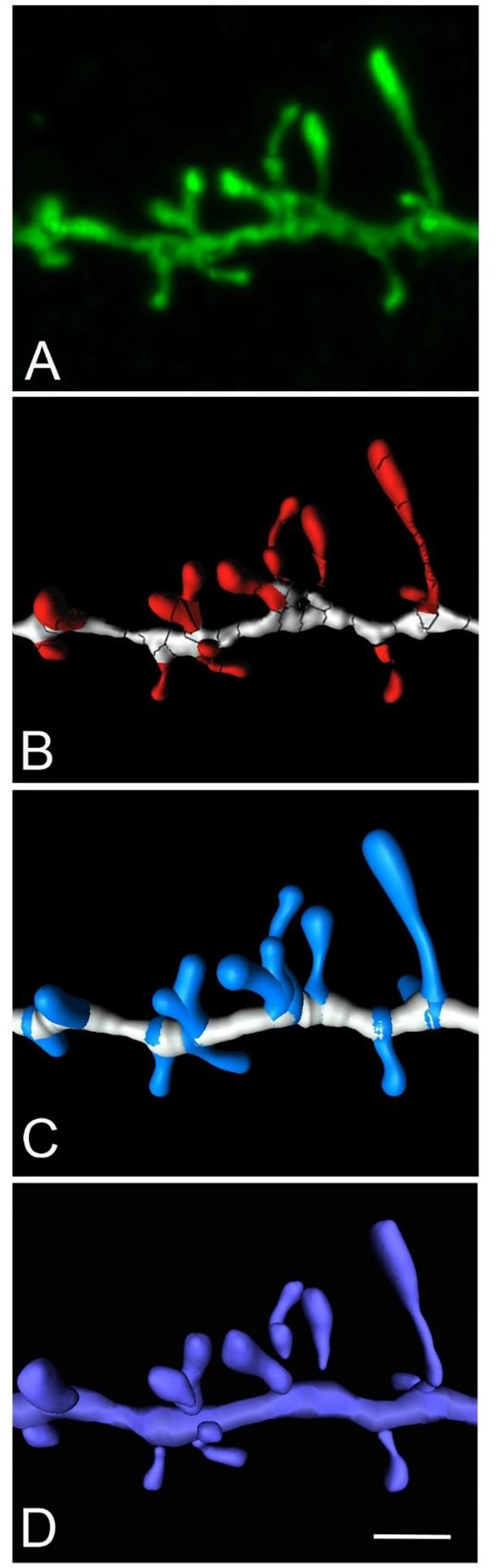
High magnification image of a human basal dendritic segment from an intracellularly injected layer III pyramidal neuron. (B,C) Three-dimensional reconstruction of the morphology of each dendritic spine shown in (A), using Imaris isosurfaces (B) and Imaris filament tracer (C). (D) The building of the spines from the reconstruction is shown in (B), using Neuronize after the unification and repair process. Scale bar (in D): 2 μm in (A–D).