
Figure 10b.
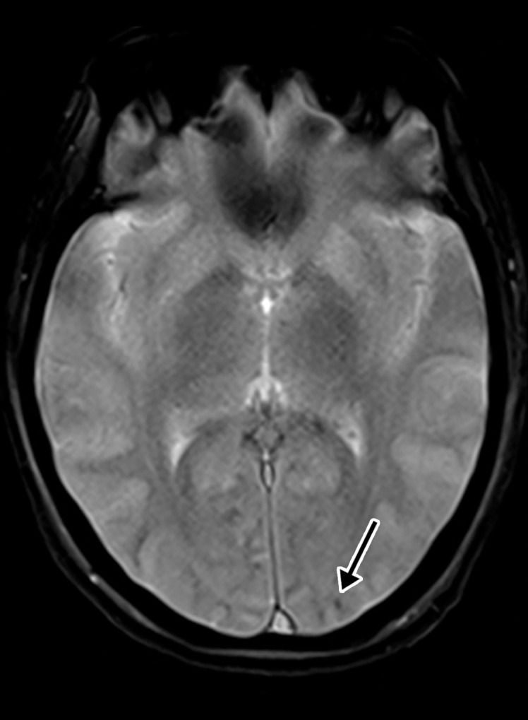
Hemorrhagic diffuse leukoencephalopathy in a 43-year-old woman with COVID-19. The hospital stay was complicated by inability to follow commands and persistently depressed status after extubation. The laboratory test results showed markedly elevated d-dimer levels (>7.6 mg/mL), international normalized ratio level (1.4), and platelet count (290). (a) Axial T2-weighted MR image shows confluent bilateral signal hyperintensities (arrows) in the white matter of both parietal lobes. No associated diffusion restriction was depicted on diffusion-weighted or ADC images (not shown). (b) Axial gradient-echo MR image shows punctate microhemorrhages (arrow) in the subcortical white matter of the left occipital lobe. Multiple microhemorrhages were scattered throughout the cerebral hemispheres bilaterally and in the splenium of the corpus callosum (not shown).