. 2020 Nov 2;40(7):1866–1892. doi: 10.1148/rg.2020200195

Figure 26c.

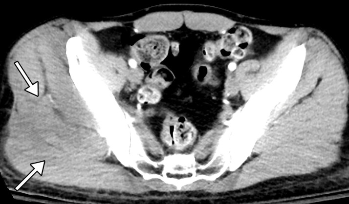
Rhabdomyolysis in a 34-year-old man who presented with minor trauma and was diagnosed with COVID-19 in the emergency department. Laboratory analysis results demonstrated an extremely high creatine kinase level of 35 145 U/L (normal range, 39–308 U/L). (a–c) Coronal (a) and axial (b, c) contrast-enhanced CT images of the pelvis show an enlarged right gluteal region (arrows) without associated fracture. (d–f) Axial (d) and coronal (e) contrast-enhanced chest CT images and a CT image with an artificial intelligence deep learning algorithm for the detection of pulmonary embolism (f) show bilateral multifocal multilobar peribronchovascular patchy airspace and ground-glass opacities (arrows in d), which are a typical finding for COVID-19. Multiple segmental and subsegmental right lower lobe pulmonary emboli (arrows in e and f) were also detected.

Rhabdomyolysis in a 34-year-old man who presented with minor trauma and was diagnosed with COVID-19 in the emergency department. Laboratory analysis results demonstrated an extremely high creatine kinase level of 35 145 U/L (normal range, 39–308 U/L). (a–c) Coronal (a) and axial (b, c) contrast-enhanced CT images of the pelvis show an enlarged right gluteal region (arrows) without associated fracture. (d–f) Axial (d) and coronal (e) contrast-enhanced chest CT images and a CT image with an artificial intelligence deep learning algorithm for the detection of pulmonary embolism (f) show bilateral multifocal multilobar peribronchovascular patchy airspace and ground-glass opacities (arrows in d), which are a typical finding for COVID-19. Multiple segmental and subsegmental right lower lobe pulmonary emboli (arrows in e and f) were also detected.