
Figure 3d.
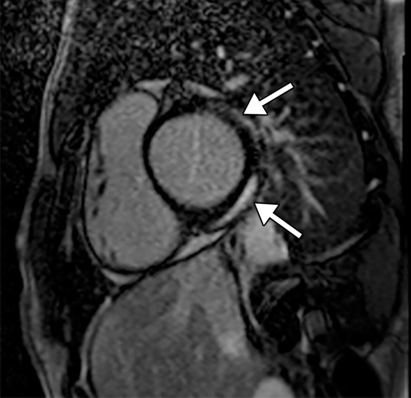
Myocarditis in a 17-year-old adolescent boy who presented to the pediatric emergency room with chest pain and was diagnosed with elevated troponin levels, diffuse ST-segment elevation at electrocardiography, ventricular ectopy, and COVID-19. (a) Cardiac short-axis T2-weighted fat-saturated black-blood MR image shows diffuse increased signal intensity in the subepicardial layer of the septal, lateral, and inferolateral walls in the basal segment (arrows) and subepicardial layer of the lateral and inferolateral walls of the midventricular segments (not shown), consistent with edema. (b) Precontrast T1-weighted short-axis fat-saturated MR image shows normal myocardial thickness and signal intensity (arrows). (c, d) Postcontrast short-axis T1-weighted cardiac MR images obtained at the same level as b show early (c) and delayed (d) subepicardial enhancement of the basal anterior, anterolateral, inferolateral, and inferior segments in a nonischemic pattern (arrows), findings consistent with myocarditis given the clinical context. The enhancement area corresponds to the T2 signal abnormality depicted in a.