. 2020 Nov 2;40(7):1866–1892. doi: 10.1148/rg.2020200195

Figure 4.

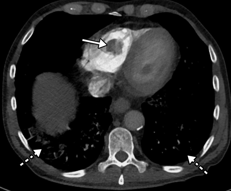
Right ventricular thrombus and COVID-19 pneumonia in a 62-year-old man with hypoxia and markedly elevated d-dimer levels (>50 000 ng/mL). Axial chest CT angiographic image shows a well-circumscribed hypoattenuating filling defect within the lumen of the right ventricle, indicative of a thrombus (solid arrow). Patchy lung opacities (dashed arrows) related to COVID-19 pneumonia are only vaguely depicted, owing to the window settings used.

Right ventricular thrombus and COVID-19 pneumonia in a 62-year-old man with hypoxia and markedly elevated d-dimer levels (>50 000 ng/mL). Axial chest CT angiographic image shows a well-circumscribed hypoattenuating filling defect within the lumen of the right ventricle, indicative of a thrombus (solid arrow). Patchy lung opacities (dashed arrows) related to COVID-19 pneumonia are only vaguely depicted, owing to the window settings used.