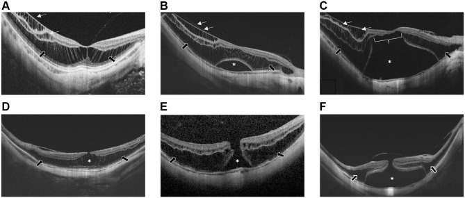
Figure 1.
(A) Foveoschisis/maculoschisis/retinoschisis (FS/MS/RS). A separation of retinal layers, which remain connected by Müller cells stretched in multiple columnar structures appears in both inner retinal layers (white arrows, inner RS) and in outer retinal layers (black arrows, outer RS). (B) Foveal detachment (FD). Asterisk indicates the FD. White arrows shows the inner RS, black arrows the outer RS. (C) Retinal detachment (RD) (asterisk) associated with inner RS (white arrows) and outer RS (black arrows). White line indicates outer lamellar macular hole (O-LMH). (D) Lamellar macular hole (LMH). Asterisk indicates the partial foveal defect with intact outer retinal layers. Black arrows show the outer RS. (E) Full-thickness macular hole (FTMH). Asterisk indicates the FTMH associated with outer RS (black arrows). (F) Full-thickness macular hole with retinal detachment (MHRD). Asterisk indicates the MHRD associated with outer RS (black arrows).

