Figure 1.
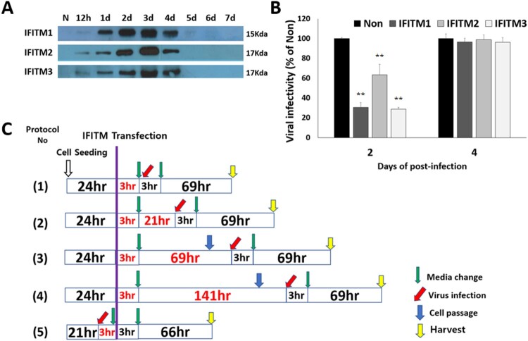
Scheme of FFV infection experiments. (A) Transient expression patterns of IFITMs. In 6-well plate, 2 × 10⁵ CRFK cells were grown for 1 day before transfection. On the day of transfection, 2.6 µg of the IFITM 1, 2, 3 expression vectors were transfected by PEI. At the indicated post-transfection days, cell lysates were harvested by centrifuging at 22,000 g for 15 min at 4°C. The collected cell lysates were stored at –20°C for later usage. Expression of IFITM1, IFITM2, and IFITM3 in all samples was confirmed by western blotting with antibodies to IFITM1 (Santa Cruz), IFITM2 (Proteintech), and IFITM3 (Abcam). (B) Initial experiments of FFV and IFITMs. In 6-well plate, 2×10⁵ CRFK cells were grown for 1 day before transfection. 2.6 µg of the IFITM 1, 2, 3 expression vectors were transfected by PEI on the next day. FFV was infected at 2 days post-transfection (MOI 1.0). The supernatants were collected at 2, 4 post-infection days, and the virus titers were measured by FeFAB assay. C. Five different protocols of FFV infection experiments. In 6-well plates, 2 × 10⁵ CRFK cells were grown for 1 day before transfection. On the day of transfection, 2.6 µg of the IFITM 1, 2, 3 expression vectors were transfected by PEI. After 3 h, the medium was exchanged for fresh medium (green arrow). FFV infection took place at 3 h after (1), 1 day after (2), 3 days after (3), 6 days after (4), and 3 h before (5) transfection. Green arrows indicate a change in medium (3 h). Red arrows indicate virus infection (MOI 1.0). Blue arrows indicate cell passaging, to prevent cell accumulation and death.

