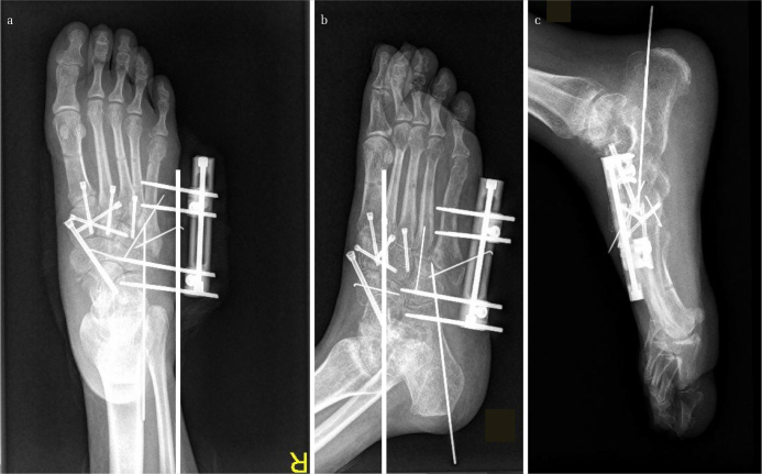
Figure 5. a–c.
Images of the foot in Figure 1 after the bilateral frame was removed, and the tarsometatarsal and the Chopart joints were anatomically reduced and fixed. Excellent reduction and fixation with cannulated screws of the medial and the middle columns could be noted. The fractured talonavicular joint was transfixed with a cannulated screw. For the comminuted lateral column, a small unilateral external fixator together with K-wires was used

